
Overview
C-SAM (Confocal Scanning Acoustic Microscopy) is a widely adopted technique in advanced manufacturing for non-destructive quality control of brazed components.
The process relies on the propagation and reflection of high-frequency ultrasound waves to reveal internal features that are otherwise invisible through surface inspection.
In brazing processes, where filler materials join intricate metallic structures, even microscopic defects can compromise performance.
Delamination, voids, clogged channels, or incomplete bonding may occur during fabrication and can significantly affect the mechanical integrity, leak tightness, or thermal performance of the final assembly.

C-SAM offers a unique capability to:
- Visualize interfaces at micrometer-scale resolution.
- Detect hidden defects such as voids, cracks, or weak bonds.
- Quantify bond uniformity and verify braze depth.
- Perform full-area scanning without destructive sectioning.
Because of its high sensitivity and precision, C-SAM has become a key quality control tool in industries where brazed joints are critical, including aerospace, energy, medical devices, precision (machine) manufacturing and (micro-)electronics.
Objective and Sample
The goal of this study was to perform a non-destructive evaluation using acoustic microscopy on parts to:
- Detect possible delamination or clogged channels.
- Assess the depth of the first interface.
- Identify potential defects at soldered joints.
Due to confidentiality constraints, exact sample images, composition and dimensions are subject to “confidential information” under NDA and cannot be disclosed.
Analysis Principle
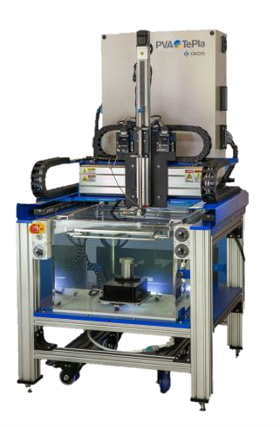
The inspection was conducted using an acoustic microscopy system in reflection mode.
A scan was performed across the full surface of the parts, with a lateral resolution in the range of 10–20μm.
The primary region of interest was the braze filler material between etched metallic plates of approximately 1 mm thickness.
The images generated during the analysis represent the amplitude of reflected ultrasonic waves, displayed in grayscale to highlight internal features and possible discontinuities.
Results
As all images and detailed results are subject to confidentiality under NDA agreement, they cannot be disclosed here.
However, the evaluation successfully provided high-resolution insight into the structural integrity of the samples, particularly at the interfaces of the brazed regions.
Conclusion
The non-destructive acoustic microscopy analysis revealed:
- No major delamination or clogging was observed across the inspected regions.
- Minor defects in the micrometer range were detected.
- The relevance of these minor irregularities must be assessed according to the internal acceptance criteria of the end-user.
This case demonstrates the effectiveness of acoustic microscopy as a powerful diagnostic tool for evaluating soldered and brazed components without causing damage, ensuring structural quality while preserving the integrity of the samples.
Reference
Instrument Manual: OKOS 2020 NDT-CF 300